Mitochondrial structureTomographical studies by Carmen Mannella |

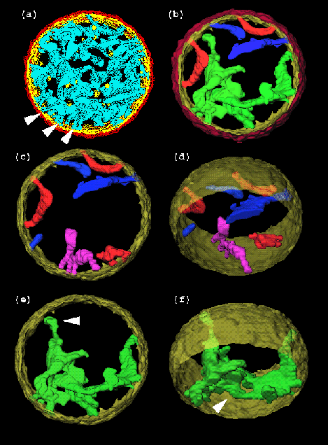
(a) Stacked contours showing the outer membrane (red), the inner membrane (periphery, yellow; cristae, blue), and matrix granules (yellow). The mitochondrion has an outer diameter of 1.5 µm. Arrows point to some of the narrow tubular connections of cristae to the periphery of the inner membrane. Contours were drawn using the Sterecon system from 1.6 nm-thick slices parallel to the plane of the tilt axis (i.e. the plane of the page).
(b - d) Surface renderings of the model in (a) showing selected cristae with one tubular connection to the inner peripheral membrane (blue), two connections (orange-red) and four connections (red-purple).
(e, f) The surface in green might represent a single crista with several interconnected compartments, or two cristae in close apposition at the arrow (f). The arrow in (e) points to a region that is over 1 µm from the nearest opening into the inner peripheral membrane.
Ref:
Mannella, C., Marko, M. and Buttle, K. (1997) Reconsidering mitochondrial structure: new views of an old organelle. TIBS 22, 37-38
(Picture kindly provided by Carmen Mannella. Legend adapted from the reference above.)